
How Digital Scans Are Changing Orthodontic Treatment
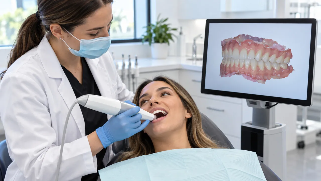
Orthodontic treatment has changed significantly with advances in dental technology. One of the most useful developments is the digital intraoral scan, which allows orthodontists to create a detailed three dimensional image of the teeth and surrounding structures without relying on traditional impression materials.
For many patients, digital scans can make the orthodontic experience more comfortable while giving the orthodontic team detailed information for treatment planning. From braces to clear aligners, digital technology is becoming an important part of modern orthodontic care.
What Are Digital Dental Scans?
A digital dental scan uses a small handheld intraoral scanner to capture images of the teeth and oral structures. The scanner is gently moved around the mouth, collecting multiple images that software combines into a three dimensional digital model.
Unlike conventional dental impressions, which use trays filled with impression material, digital scanning does not require the patient to hold a large amount of material in the mouth while it sets.
The resulting digital model can be viewed on a computer and used as part of the orthodontic assessment and treatment planning process.
Why Are Digital Scans Useful In Orthodontics?
Orthodontic treatment depends on understanding how the teeth fit together and how they are positioned within the dental arches. A digital scan provides a detailed visual representation that can help the orthodontist assess these relationships.
Digital models can be useful for evaluating:
- Tooth position and alignment
- Dental arch shape
- Crowding and spacing
- Bite relationships
- Changes during orthodontic treatment
- Progress toward the planned result
The scan is one part of a comprehensive orthodontic assessment and may be combined with clinical examinations, photographs, and dental X rays when needed.
Are Digital Scans More Comfortable Than Traditional Impressions?
For many patients, comfort is one of the biggest advantages of digital scanning.
Traditional impressions require a tray and impression material that can sometimes trigger a gag reflex or create a feeling of pressure in the mouth. Digital scanning eliminates the need for that material.
The scanner is moved gradually around the teeth, allowing the orthodontist or dental professional to capture the necessary images. The process is generally straightforward and does not involve setting impression material.
This can be particularly helpful for patients who have difficulty tolerating conventional impressions.
How Do Digital Scans Help With Clear Aligners?
Digital scanning has become especially useful in clear aligner treatment. A digital model can be used to plan tooth movements and develop a series of aligners designed around the patient's treatment goals.
Depending on the orthodontic system being used, digital records can also help visualize proposed tooth movements before treatment begins.
However, digital scans do not determine whether clear aligners are suitable on their own. The orthodontist still needs to evaluate the patient's teeth, bite, jaw relationship, oral health, and overall treatment needs.
Can Digital Scans Help Track Treatment Progress?
Yes. New digital scans can be taken during treatment and compared with earlier records.
This can help the orthodontic team evaluate changes in tooth position and determine whether treatment is progressing as expected. Digital records may also make it easier to visualize changes that can be difficult to appreciate from a quick visual examination alone.
For patients, seeing changes in a digital model can make the progress of treatment easier to understand.
How Do Digital Scans Improve Treatment Planning?
One of the major benefits of digital orthodontics is the ability to work with a detailed digital model rather than a physical impression.
Digital records can be stored electronically and accessed when needed. They can also be integrated into certain orthodontic planning systems and laboratory workflows.
Depending on the treatment, digital scans may support the planning of:
- Clear aligners
- Orthodontic appliances
- Braces
- Retainers
- Other customized orthodontic treatments
Digital planning can also help improve communication between the orthodontist, dental laboratory, and other members of the treatment team when collaboration is required.
Can Patients See Their Digital Scan?
In many practices, patients can view their digital dental model on a screen. This can make it easier to understand areas of crowding, spacing, bite problems, or tooth positioning.
Visualizing the condition of the teeth may help patients better understand why a particular treatment has been recommended and what the orthodontist is trying to achieve.
A digital model is not a prediction of exactly how every patient's treatment will progress, but it can be a valuable educational and planning tool.
Are Digital Scans Accurate?
Modern intraoral scanners can provide highly detailed digital models that are useful for many orthodontic applications. However, scan quality depends on the scanner, scanning technique, the patient's ability to remain still, and the conditions inside the mouth.
Saliva, movement, limited access, and other factors can sometimes affect image capture. The orthodontic team reviews the digital record to make sure it contains the information needed for treatment planning.
Digital scanning also does not replace other diagnostic tools when those tools are clinically necessary.
Do Digital Scans Replace Dental X Rays?
No. Digital scans and dental X rays provide different types of information.
An intraoral scan primarily shows the visible surfaces and shape of the teeth and dental arches. X rays can provide information about structures that cannot be seen directly, such as tooth roots and the surrounding bone.
Depending on the patient's age, symptoms, dental development, and treatment needs, an orthodontist may recommend X rays or other diagnostic imaging in addition to digital scans.
What Does A Digital Orthodontic Scan Appointment Feel Like?
The scanner is typically moved around the mouth in a controlled manner while it captures images of the teeth. There is no impression material that needs to set.
The length of the appointment can vary depending on the scanner, the patient's mouth, and whether both arches and the bite need to be recorded.
Once the scan is complete, the orthodontic team can review the digital model and use it as part of the patient's orthodontic records.
Are Digital Scans Suitable For Everyone?
Digital scanning can be used for many children, teenagers, and adults, but the appropriate diagnostic approach depends on the individual.
Some patients may require additional examinations or imaging before treatment can be planned. The orthodontist will determine which records are necessary based on the patient's specific dental and orthodontic needs.
A More Comfortable Approach To Modern Orthodontics
Digital scanning is making orthodontic records more convenient, comfortable, and interactive for many patients. By creating detailed three dimensional models, this technology can support diagnosis, treatment planning, progress monitoring, and communication throughout orthodontic care.
If you are considering orthodontic treatment, Dr. Parijat Pallav can assess your teeth and bite and explain how digital technology may be incorporated into your personalized treatment plan.
FAQs
Q: Are digital dental scans painful?
A: No. Digital scanning is generally noninvasive and does not involve impression material. The scanner is gently moved around the teeth to capture images.
Q: How long does an orthodontic digital scan take?
A: The scanning process can vary depending on the patient and the type of records required. Your orthodontic team can explain how long the appointment is expected to take.
Q: Can digital scans be used for clear aligners?
A: Yes. Digital scans are commonly used to create digital models that can support clear aligner treatment planning and fabrication.
Q: Do digital scans replace X rays?
A: No. Digital scans and X rays provide different information. Your orthodontist may recommend both when necessary for proper diagnosis and treatment planning.
Q: Can digital scans show orthodontic treatment progress?
A: Yes. Digital scans taken at different stages can be compared to assess changes in tooth position and treatment progress.
Our Location

)

